


Morphology of the Neurocranium after COMPAGNO, 1988
Material: Neurocranium of a triakid. Gogolia filewoodi, CAS-27588, 224 mm. fetus, to show external terminology of a typical carcharhinoid cranium. A. Dorsal. B. Ventral. C. Lateral views. Figure after Compagno (1973d), lettering of some abbreviations different than elsewhere in this work equivalents given where necessary) (Ref. Compagno, 1988, image redrawn)
I. Dorsal view:
AF = anterior fontanelle
ASC = anterior semicircular canal
FM = foramen magnum
FEN = endolymphatic foramen
FOE and FOI = external and orbital foramina of the preorbital canal (= PC) transmitting the superficial ophthalmic (parts of V, trigeminal and VII, facial) nerve through the supraorbital crest
FPE and FPI = external and orbital profundus foramina ( = PF), transmitting the deep ophthalmic nerve through base of preorbital process
FPN = perilymphatic foramen
LR = lateral rostral cartilages
NC = nasal capsule
OT = otic capsule
PR = preorbital process
PRF = parietal fossa
PSC = posterior semicircular canal
PT = postorbital process
RF = rostral fenestra
RRF = ridge connecting base of lateral rostral cartilage with edge of anterior fontanelle
SC = supraorbital crest
SR = sphenoptcrotic ridge
SS = suborbital shelf
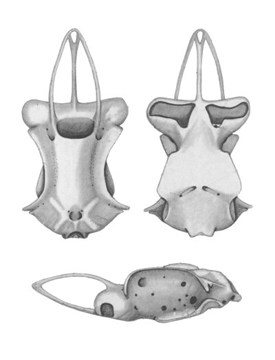


II. Ventral view:
AFV (= FSM) = foramen in subnasal membrane between facial and nasal sinuses
BP = basal plate
ECN = ectethmoid condyle
FC (=CF) = foramen for internal carotid artery
FS (= SF) = stapedial foramina for stapedial (orbital) artery extending through suborbital shelf, with both basal and orbital foramina shown
HF = hyomandibular facet
MR = medial rostral cartilage
NA = nasal aperture
NF = nasal fontanelle
NP = orbital notch
OC = occipital condyle
OCN = occipital centrum
OR = opisthotic ridge
RN = rostral node
SEF = subethmoid fossa
SS = suborbital shelf


III. Lateral view:
ECN = ectethmoid condyle
FCV = foramen for anterior cerebral vein
FES = foramen for efferent spiracular (pseudobranchial) artery
FOC = foramen for superficial ophthalmic nerve through medial wall of orbit into cranial cavity
FOE and FOI = external and orbital foramina of the preorbital canal (= PC) transmitting the superficial ophthalmic (parts of V, trigeminal and VII, facial) nerve through the supraorbital crest
FPC = foramen for deep ophthalmic (profundus, part of V, trigeminal) nerve through medial wall of orbit into cranial cavity
FPE and FPI = external and orbital profundus foramina ( = PF), transmitting the deep ophthalmic nerve through base of preorbital process
FS (= SF) = stapedial foramina for stapedial (orbital) artery extending through suborbital shelf, with both basal and orbital foramina shown
F II = optic nerve foramen
F III = oculomotor nerve foramen
F IV = trochlear nerve foramen
F IX (= FG) = glossopharyngeal nerve foramen
F X (= FV) = vagus nerve foramen
HF = hyomandibular facet
IOC = interorbital canal for posterior cerebral vein
LR = lateral rostral cartilages
MR = medial rostral cartilage
NA = nasal aperture
NC = nasal capsule
NP = orbital notch
O = orbit
OC = occipital condyle
ONF (= OF) = orbitonasal foramen
OR = opisthotic ridge
ORF = orbital fissure (trigeminofacialis chamber), through medial wall of orbit for major branches of trigeminal (V) and facial (VII) nerves
OT = otic capsule
PT = postorbital process
RN = rostral node
RRF = ridge connecting base of lateral rostral cartilage with edge of anterior fontanelle
SC = supraorbital crest
SR = sphenoptcrotic ridge
SS = suborbital shelf