


Mennerotodus parmleyi
Cicimurri, Ebersole & Martin, 2020
Classification: Elasmobranchii Lamniformes Odontaspididae
Reference of the original description
Two new species of Mennerotodus Zhelezko, 1994 (Chondrichthyes: Lamniformes: Odontaspididae), from the Paleogene of the southeastern United States. Fossil Record, 23(2), 117–140
Two new species of Mennerotodus Zhelezko, 1994 (Chondrichthyes: Lamniformes: Odontaspididae), from the Paleogene of the southeastern United States. Fossil Record, 23(2), 117–140
Synonyms / new combinations and misspellings
Mennerotodus cf. parmleyi
Mennerotodus cf. parmleyi
Types
Mennerotodus parmleyi
Holotype: SC: 2004.34.175; Paratype: SC: 2013.44.117; SC: 2013.44.119; SC: 2013.44.122; SC: 2013.44.120; SC: 2013.44.128; SC: 2013.44.130; SC: 2013.44.132; SC: 2004.34.182; SC: 2013.44.157; SC: 2004.34.181;
Mennerotodus parmleyi
Holotype: SC: 2004.34.175; Paratype: SC: 2013.44.117; SC: 2013.44.119; SC: 2013.44.122; SC: 2013.44.120; SC: 2013.44.128; SC: 2013.44.130; SC: 2013.44.132; SC: 2004.34.182; SC: 2013.44.157; SC: 2004.34.181;
Images of types
Description:
Citation: Mennerotodus parmleyi Cicimurri, Ebersole & Martin, 2020: In: Database of fossil elasmobranch teeth www.shark-references.com, World Wide Web electronic publication, Version 07/2026
Please send your images of "Mennerotodus parmleyi" to info@shark-references.com
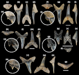
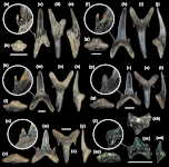
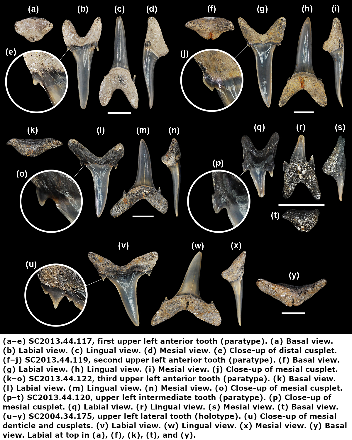
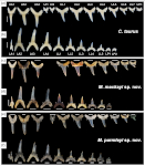
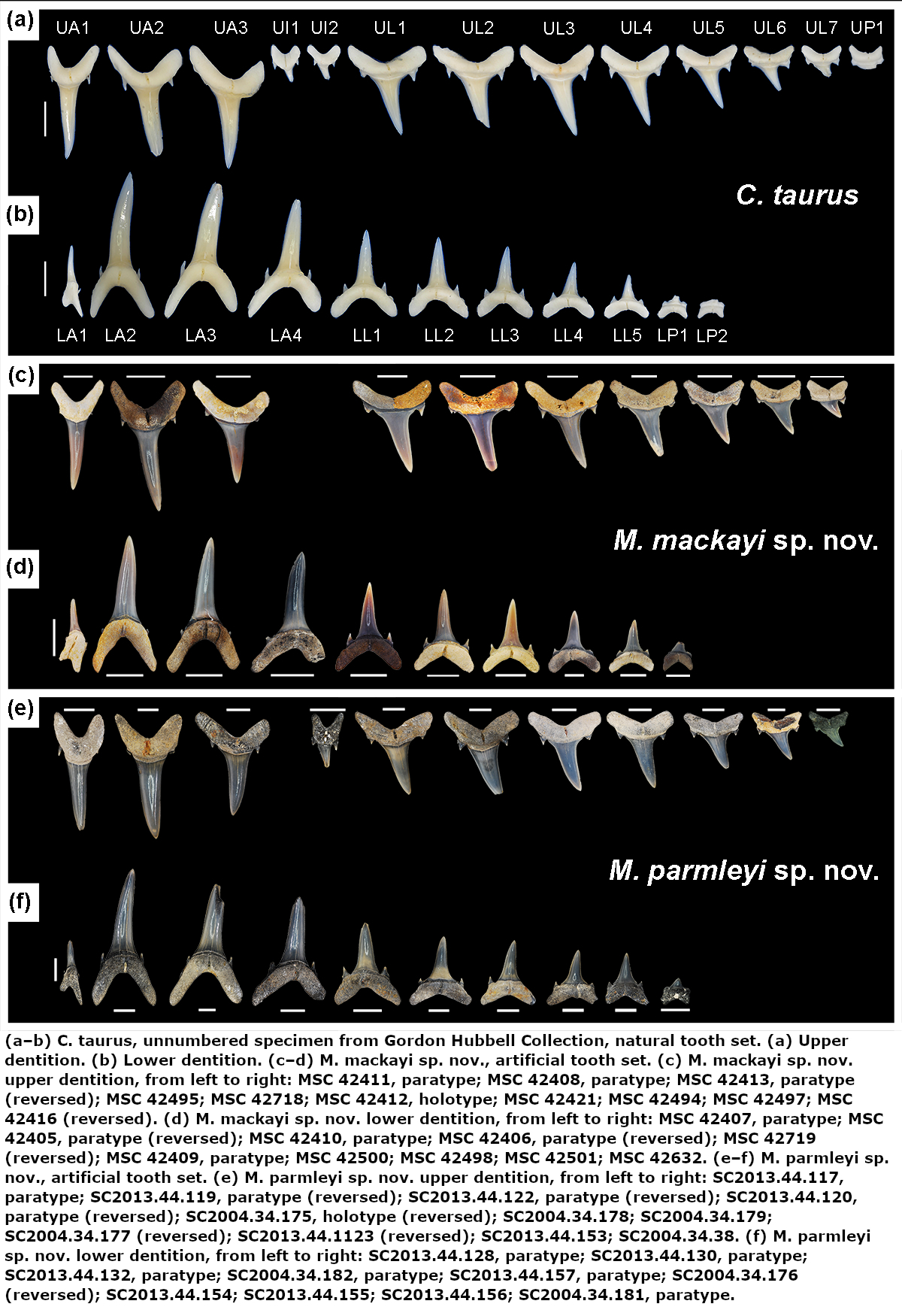
Mennerotodus parmleyi Cicimurri, Ebersole & Martin, 2020; types, Eocene (Bartonian) Clinchfield Formation, Hardie Mine, Wilkinson County, Georgia, USA (caption see images of types). Scale bars = 5 mm.
Mennerotodus parmleyi Cicimurri, Ebersole & Martin, 2020; types, Eocene (Bartonian) Clinchfield Formation, Hardie Mine, Wilkinson County, Georgia, USA (caption see images of types). Scale bars = 5 mm.
Description
Original description after Cicimurri, Ebersole & Martin (2020) [28668]:
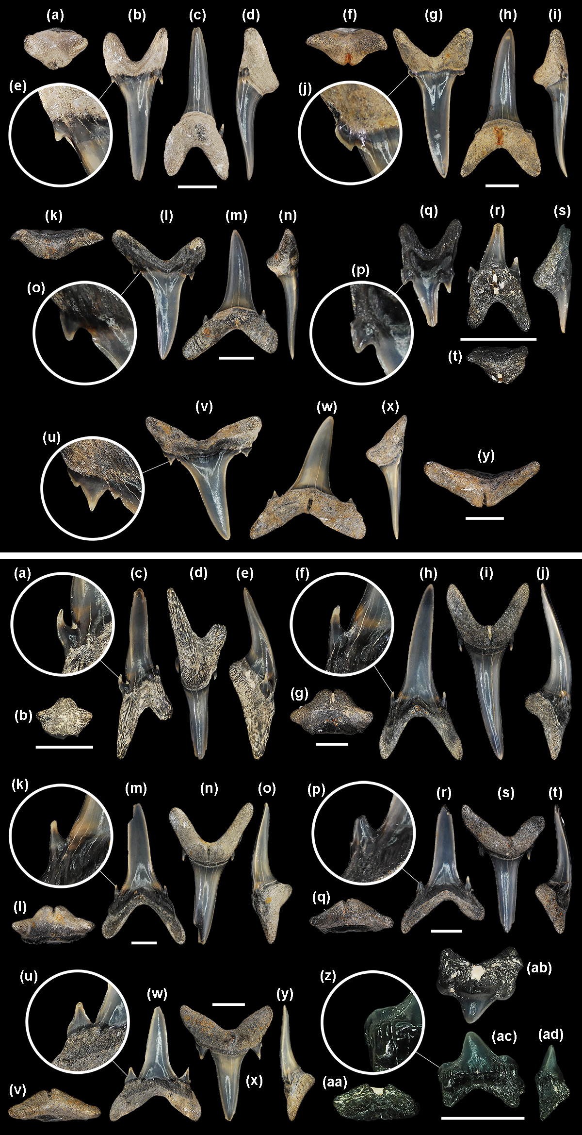
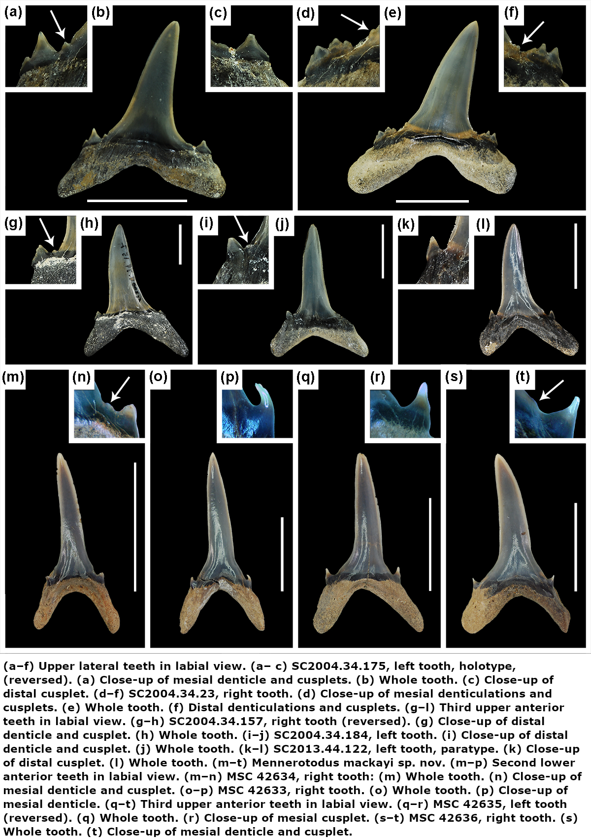
First upper anterior teeth. These small teeth, which do not exceed 21mm in total height, are slightly asymmetrical in labial view (Fig. 5b). The main cusp is very narrow, is slightly distally inclined, and has a sigmoidal profile (Fig. 5c, d). Mesial and distal cutting edges are sharp, smooth, and subparallel but never reach the base of the main cusp. There may be a minuscule tubercle or very short and sharp ridge at the very base of the main cusp, well separated from the main cutting edge (Fig. 5e). A single pair of short, conical cusplets is located at the crown foot (Fig. 5a). The labial face of the main cusp is smooth, is flat apically but weakly convex on its lower half, and in distal view appears to have a slight twist. In contrast, the lingual face is very convex and may bear faint longitudinal ridges on the lower half. The root is bilobate and has a large lingual boss that is bisected by an elongate and deep nutritive groove (Fig. 5a, c, d). The lingual dental band at the crown foot is conspicuous and sometimes deeply impressed. Root lobes are rather short and may be cylindrical or mesiodistally compressed. The distal lobe is more elongated and more obviously angled away from the nutritive groove (Fig. 5b, c).
Second upper anterior teeth. The largest specimen measures 33mm in total height. The main cusp is tall and narrow, more triangular in appearance than the main cusp of the first anterior tooth, and slightly distally inclined. Cutting edges are biconvex apically but otherwise subparallel (Fig. 5g, h), and they do not reach the cusp base (Fig. 5j). The labial face is smooth and flat to very weakly convex, but the lingual face is very convex and may bear fine vertical ridges on the lower half. The main cusp is flanked by a single pair of cusplets, although a second diminutive cusplet was occasionally observed on the mesial side (Fig. 5g, h). Cusplets are conical to triangular, sharply pointed, and lingually curved. Conical cusplets lack cutting edges, but more triangular cusplets exhibit complete cutting edges. The lingual boss bears a thin nutritive groove, and the dental band may be impressed (Fig. 5f, h). Although root lobes are of nearly the same length, the mesial lobe is labiolingually thick, mesiodistally thin, and pointed basally, whereas the distal lobe is labiolingually thin, mesiodistally wide, and rounded basally (Fig. 5g, h).
Third upper anterior teeth. The largest specimen measures 32mm in total height. Teeth from this position differ from those of the other anterior positions in having a main cusp that is distally directed, often mesially curving, and only weakly sigmoidal in profile (Fig. 5l–n). In addition, root lobes are asymmetrically developed, with the mesial lobe being much more elongated than the distal one, as well as sharply divergent from the nutritive groove (Fig. 5l, m). The labial face of the main cusp is smooth and very nearly flat, whereas the lingual face is convex and may bear very fine and discontinuous vertical ridges on the lower half. The cutting edges are smooth and sharp and extend to the crown foot (Fig. 5o). The base of the cutting edge may be continuous or denticulated. Lateral cusplets are small but broad, more labiolingually compressed, and with a more conspicuous cutting edge than those of the first two anterior files (Fig. 5l, m). The lingual dental band is conspicuous and may be impressed, and although the nutritive groove is elongated, the boss is less robust than is seen on the other two anterior files (Fig. 5k, m, n).
Intermediate teeth. A single left intermediate tooth is represented. It measures nearly 8mm in total height and 4mm in width. The crown consists of a rather short and narrow main cusp that is straight and flat in profile (not sigmoid) and slightly distally inclined (Fig. 5q–s). There is a single pair of lateral cusplets, with the distal cusplet being larger (Fig. 5q). The labial face of the main cusp is flat, whereas the lingual face is very convex, and the cutting edge is continuous from the apex to the lateral cusplets. The root is bilobate with short (the distal lobe is longer), divergent lobes having rounded ends (Fig. 5r). A large lingual boss is bisected by an elongated nutritive groove (Fig. 5r–t).
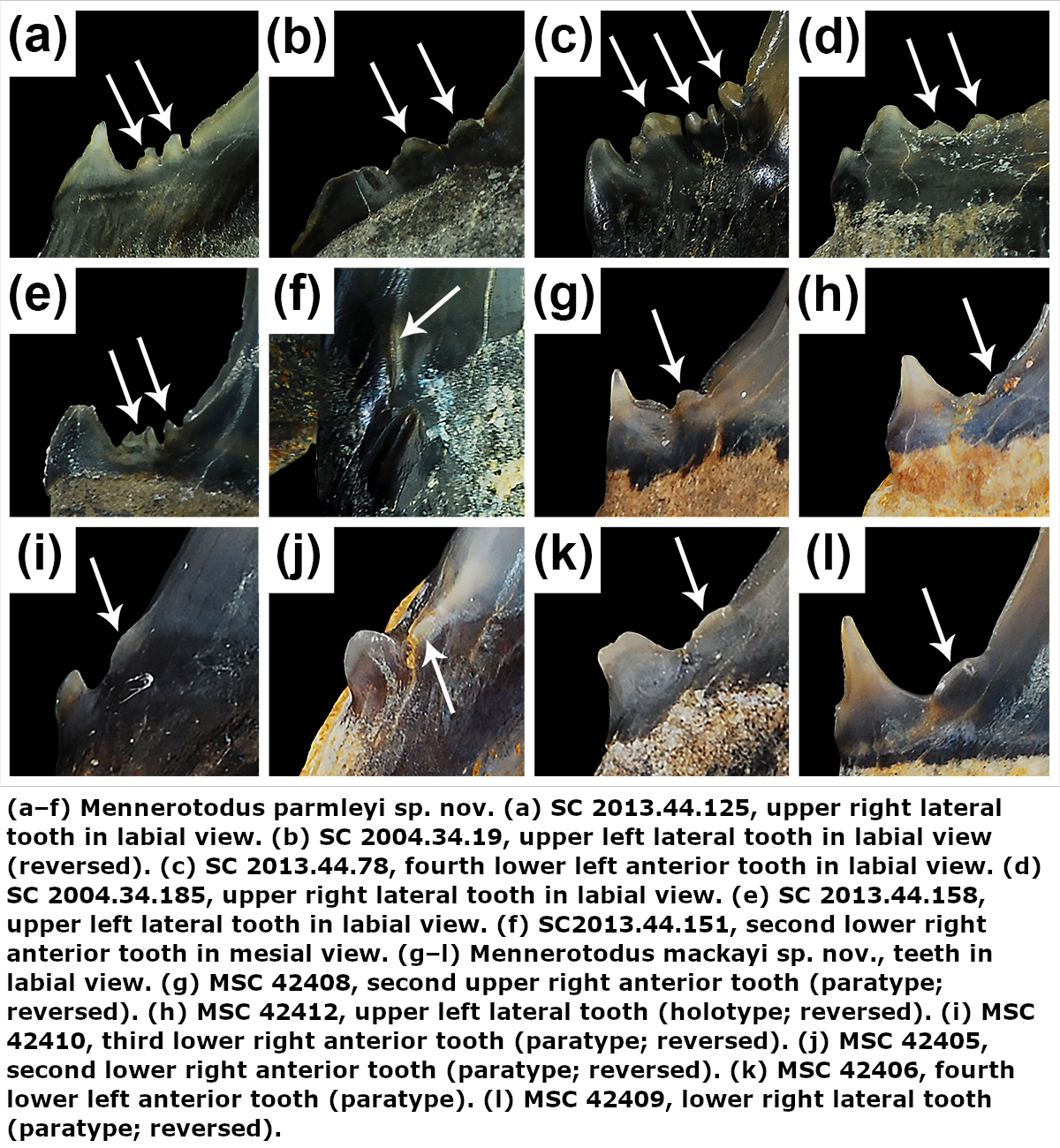
Upper lateral teeth. Upper lateral teeth can be differentiated from the anterior teeth in that the main cusp is labiolingually thinner, flat, and distally inclined, and the base is broader (Fig. 5v–x). Root lobes are shorter but wider, and they are more strongly divergent. The first few upper lateral tooth files are identified by their more elongated, narrower, and basally pointed mesial lobe, compared to the short, wide, rounded distal lobe (Fig. 5v, w). Other lateral teeth have more equidimensional root lobes and are difficult to place into a specific file. Within our sample of upper lateral teeth, it appears that the crown decreases in size but becomes more strongly distally inclined towards the commissure. The main cusp of lateral teeth is broad-based but sharply tapering, distally inclined, and straight in profile view. The labial face is smooth and flat to very weakly convex, but the lingual face is convex (although not as strongly as anterior teeth) and may bear fine vertical ridges on the lower half. The cutting edges are smooth and sharp and extend to the very base of the main cusp. The mesial and distal cutting edges may be straight, but more commonly the upper part of the main cusp appears distally curving because the mesial edge is convex and the distal edge straight to concave (Fig. 5v). The base of the cutting edge may be continuous and sharp or sometimes punctuated by one or more rounded-to-pointed denticles (Fig. 5u). The main cusp is usually flanked by a single pair of low, broadly triangular lateral cusplets (Fig. 5v, w), but occasionally a poorly developed second pair was observed (Fig. 5u). The lingual face of each cusplet is more convex than the labial face, and the cutting edge is complete from the mesial to distal side. An elongated and deep lingual nutritive groove divides the root into roughly equidimensional, subrectangular lobes with pointed ends (Fig. 5w, y). The interlobe area is V-shaped, and many teeth have a labiobasal depression at the crown base. The lingual dental band is impressed, but the root boss is less distinctive than on anterior teeth (Fig. 5y). Root width is nearly equal to total tooth height.
Upper posterior teeth. No upper posterior teeth have been identified in the sample.
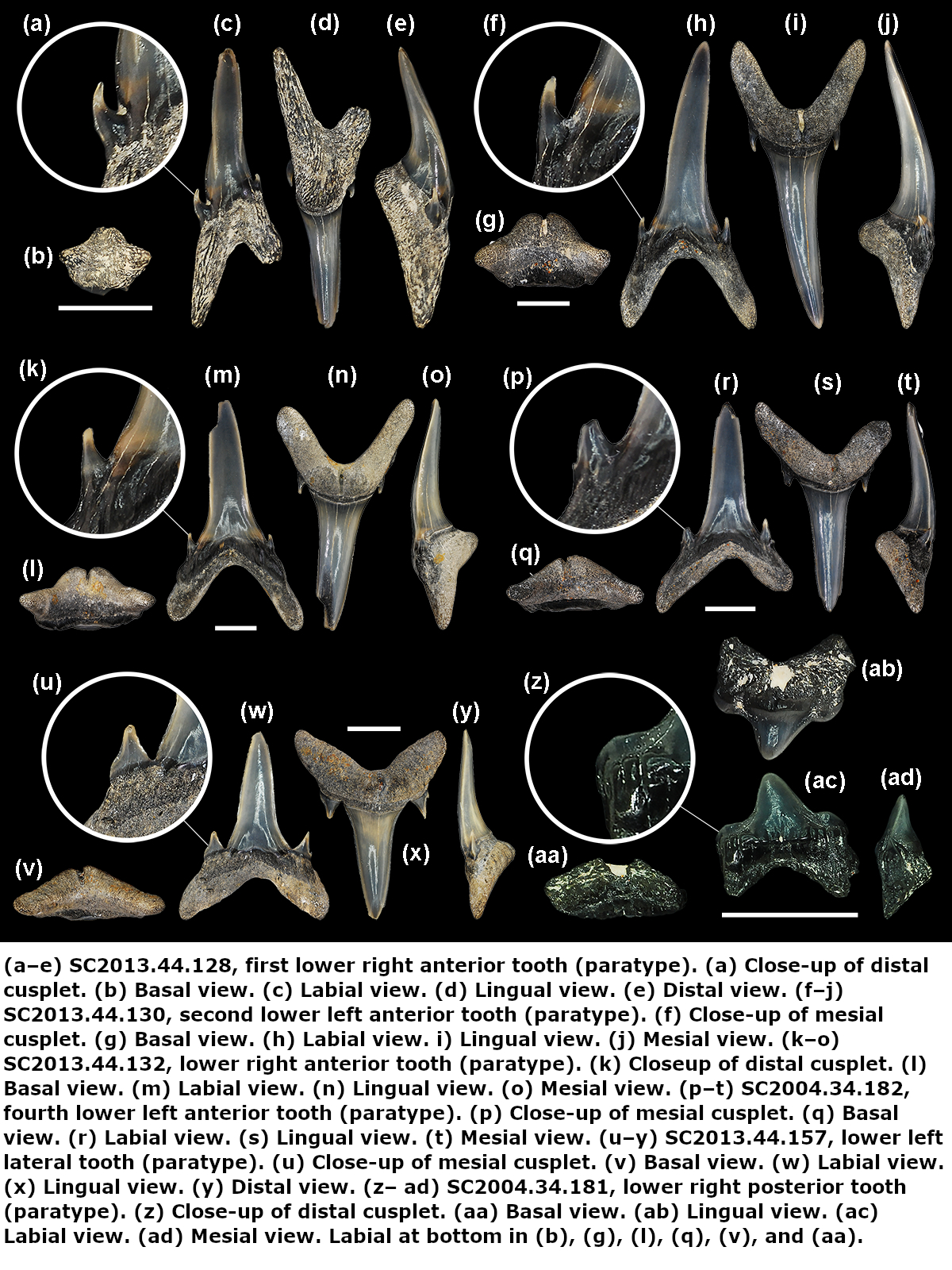
First lower anterior teeth. Teeth from this file are not known to exceed 13mmin total height. The main cusp is very narrow and may be straight to weakly curved distally, and it is inclined towards the symphysis (Fig. 6c, d). The labial face is weakly convex and smooth, whereas the lingual face is very convex (Fig. 6e). Cutting edges are smooth, sharp, and subparallel and do not reach the crown foot. A minuscule tubercle or very short and sharp edge, well separated from the main cutting edge, may occur. A single pair of lateral cusplets flanks the main cusp, and in labial view the mesial cusplet appears to be located higher on the tooth than the distal cusplet (Fig. 6c, d). Cusplets are small, conical, sharply pointed, and lingually curving (Fig. 6a). The root is laterally compressed and bilobate with a much shorter mesial lobe, and the large lingual boss is bisected by a nutritive groove (Fig. 6b–e). Root height is equal to crown height.
Second lower anterior teeth. Teeth in this position are symmetrical and reach at least 32mm in total height. The main cusp is tall, narrow, and erect and has a sigmoidal profile (Fig. 6h–j). The labial face is smooth and flat apically but may be more convex near the base, whereas the lingual face is very convex (Fig. 6j) and may bear fine vertical ridges on the lower half. The cutting edges are subparallel and appear biconvex due to medial curvature near their base, and the edges end well before the cusp base (Fig. 6h, i). A single pair of conical, sharply pointed, and lingually curved lateral cusplets is present (Fig. 6f). The root is bilobate with a large lingual boss that is bisected by a deep nutritive groove, and the dental band is wide and impressed (Fig. 6g, i, j). Root lobes are elongated and of equal length, although the mesial lobe may be slightly wider (Fig. 6h, i). Root height is roughly one-third (30 %) of the total tooth height.
Third lower anterior teeth. These teeth (Fig. 6k–o) are essentially the same as those of the second anterior file. However, they can be distinguished by their more divergent root lobes and an elongated and narrower mesial root lobe compared to the distal lobe (Fig. 6m, n).
Fourth lower anterior teeth. Teeth in this file (Fig. 6p–t) are morphologically similar to those in the third upper anterior file (i.e., Fig. 5k–o), but they differ in that the main cusp is distally inclined but not mesially curving and both root lobes are slightly more elongated (Fig. 6r, s). Cutting edges very nearly reach the crown foot. A single pair of lateral cusplets flanks the main cusp, and these cusplets bear conspicuous cutting edges and are broader than those on the more proximal anterior teeth (Fig. 6p–s). The distal root lobe is shorter, wider, and more rounded than the mesial lobe, which is elongated and pointed at the end (Fig. 6r, s). The root lobes of teeth from this file are more divergent than on the third lower anterior file (compare Fig. 6m to Fig. 6r).
Lower lateral teeth. Lower lateral teeth can be differentiated from the anterior teeth in that the crown is shorter, labiolingually thinner, and rather flat and cutting edges extend to the crown foot (Fig. 6w–y). Root lobes are shorter but wider, and they are more widely separated (Fig. 6x). The root lobes in the first few lower lateral files have a slightly shorter and wider mesial lobe compared to the distal lobe, but other lateral teeth have more equidimensional root lobes and are difficult to place into a specific file. Within our sample of lower lateral teeth, it appears that the crown decreases in size and becomes slightly distally inclined towards the commissure. Lower lateral teeth are distinguished from upper lateral teeth by having erect main cusps as opposed to conspicuously distally inclined ones, and root lobes are shorter, lower, and more pointed (compare Fig. 6w to Fig. 5v). In general, the main cusp is broad-based but sharply tapering, vertical to slightly distally inclined, and virtually flat with little to no lingual curvature. The labial face is smooth and flat to very weakly convex, but the lingual face is convex (although not as strongly as on anterior teeth) and may bear fine vertical ridges on the lower half. The cutting edges are smooth and sharp and extend to the very base of the main cusp (Fig. 6u). The mesial and distal cutting edges are usually straight, but some teeth exhibit a mesial edge that is convex on its upper part. The base of the cutting edge may be smooth and continuous or punctuated by one or more rounded-topointed denticles. The main cusp is flanked by a single pair of low, broadly triangular lateral cusplets (Fig. 6w, v), but a poorly developed second pair on one or both sides sometimes occurs. The lingual face of the cusplet is more convex than the labial face, and the cutting edge is complete from the mesial to distal side. An elongated and deep lingual nutritive groove divides the root into low, roughly equidimensional lobes with rounded or pointed ends (Fig. 6w, x). The lingual dental band is impressed, but the root boss is less distinctive than on anterior teeth (Fig. 6v, y). Root width nearly equals total tooth height.
Lower posterior teeth. A single lower posterior tooth measures 5mm in height and 5.5mm in width. The crown is very low, broadly triangular, bluntly pointed, and slightly distally directed (Fig. 6ab, ac). There is a single pair of rather large but low, broad, and blunt lateral cusplets (Fig. 6z, ac). The labial crown face is flat and bears heavy basal vertical wrinkling. The root is bilobate and bisected by a lingual nutritive groove (Fig. 6aa, ac). Lobes are short, wide, and basally pointed, separated by a V-shaped interlobe area (Fig. 6ab, ac). Posterior teeth having a very low, convex crown that is poorly differentiated from the lateral cusplets, like those occurring near the jaw commissure of extant Carcharias taurus, are unknown for M. parmleyi sp. nov.
Original description after Cicimurri, Ebersole & Martin (2020) [28668]:
First upper anterior teeth. These small teeth, which do not exceed 21mm in total height, are slightly asymmetrical in labial view (Fig. 5b). The main cusp is very narrow, is slightly distally inclined, and has a sigmoidal profile (Fig. 5c, d). Mesial and distal cutting edges are sharp, smooth, and subparallel but never reach the base of the main cusp. There may be a minuscule tubercle or very short and sharp ridge at the very base of the main cusp, well separated from the main cutting edge (Fig. 5e). A single pair of short, conical cusplets is located at the crown foot (Fig. 5a). The labial face of the main cusp is smooth, is flat apically but weakly convex on its lower half, and in distal view appears to have a slight twist. In contrast, the lingual face is very convex and may bear faint longitudinal ridges on the lower half. The root is bilobate and has a large lingual boss that is bisected by an elongate and deep nutritive groove (Fig. 5a, c, d). The lingual dental band at the crown foot is conspicuous and sometimes deeply impressed. Root lobes are rather short and may be cylindrical or mesiodistally compressed. The distal lobe is more elongated and more obviously angled away from the nutritive groove (Fig. 5b, c).
Second upper anterior teeth. The largest specimen measures 33mm in total height. The main cusp is tall and narrow, more triangular in appearance than the main cusp of the first anterior tooth, and slightly distally inclined. Cutting edges are biconvex apically but otherwise subparallel (Fig. 5g, h), and they do not reach the cusp base (Fig. 5j). The labial face is smooth and flat to very weakly convex, but the lingual face is very convex and may bear fine vertical ridges on the lower half. The main cusp is flanked by a single pair of cusplets, although a second diminutive cusplet was occasionally observed on the mesial side (Fig. 5g, h). Cusplets are conical to triangular, sharply pointed, and lingually curved. Conical cusplets lack cutting edges, but more triangular cusplets exhibit complete cutting edges. The lingual boss bears a thin nutritive groove, and the dental band may be impressed (Fig. 5f, h). Although root lobes are of nearly the same length, the mesial lobe is labiolingually thick, mesiodistally thin, and pointed basally, whereas the distal lobe is labiolingually thin, mesiodistally wide, and rounded basally (Fig. 5g, h).
Third upper anterior teeth. The largest specimen measures 32mm in total height. Teeth from this position differ from those of the other anterior positions in having a main cusp that is distally directed, often mesially curving, and only weakly sigmoidal in profile (Fig. 5l–n). In addition, root lobes are asymmetrically developed, with the mesial lobe being much more elongated than the distal one, as well as sharply divergent from the nutritive groove (Fig. 5l, m). The labial face of the main cusp is smooth and very nearly flat, whereas the lingual face is convex and may bear very fine and discontinuous vertical ridges on the lower half. The cutting edges are smooth and sharp and extend to the crown foot (Fig. 5o). The base of the cutting edge may be continuous or denticulated. Lateral cusplets are small but broad, more labiolingually compressed, and with a more conspicuous cutting edge than those of the first two anterior files (Fig. 5l, m). The lingual dental band is conspicuous and may be impressed, and although the nutritive groove is elongated, the boss is less robust than is seen on the other two anterior files (Fig. 5k, m, n).
Intermediate teeth. A single left intermediate tooth is represented. It measures nearly 8mm in total height and 4mm in width. The crown consists of a rather short and narrow main cusp that is straight and flat in profile (not sigmoid) and slightly distally inclined (Fig. 5q–s). There is a single pair of lateral cusplets, with the distal cusplet being larger (Fig. 5q). The labial face of the main cusp is flat, whereas the lingual face is very convex, and the cutting edge is continuous from the apex to the lateral cusplets. The root is bilobate with short (the distal lobe is longer), divergent lobes having rounded ends (Fig. 5r). A large lingual boss is bisected by an elongated nutritive groove (Fig. 5r–t).
Upper lateral teeth. Upper lateral teeth can be differentiated from the anterior teeth in that the main cusp is labiolingually thinner, flat, and distally inclined, and the base is broader (Fig. 5v–x). Root lobes are shorter but wider, and they are more strongly divergent. The first few upper lateral tooth files are identified by their more elongated, narrower, and basally pointed mesial lobe, compared to the short, wide, rounded distal lobe (Fig. 5v, w). Other lateral teeth have more equidimensional root lobes and are difficult to place into a specific file. Within our sample of upper lateral teeth, it appears that the crown decreases in size but becomes more strongly distally inclined towards the commissure. The main cusp of lateral teeth is broad-based but sharply tapering, distally inclined, and straight in profile view. The labial face is smooth and flat to very weakly convex, but the lingual face is convex (although not as strongly as anterior teeth) and may bear fine vertical ridges on the lower half. The cutting edges are smooth and sharp and extend to the very base of the main cusp. The mesial and distal cutting edges may be straight, but more commonly the upper part of the main cusp appears distally curving because the mesial edge is convex and the distal edge straight to concave (Fig. 5v). The base of the cutting edge may be continuous and sharp or sometimes punctuated by one or more rounded-to-pointed denticles (Fig. 5u). The main cusp is usually flanked by a single pair of low, broadly triangular lateral cusplets (Fig. 5v, w), but occasionally a poorly developed second pair was observed (Fig. 5u). The lingual face of each cusplet is more convex than the labial face, and the cutting edge is complete from the mesial to distal side. An elongated and deep lingual nutritive groove divides the root into roughly equidimensional, subrectangular lobes with pointed ends (Fig. 5w, y). The interlobe area is V-shaped, and many teeth have a labiobasal depression at the crown base. The lingual dental band is impressed, but the root boss is less distinctive than on anterior teeth (Fig. 5y). Root width is nearly equal to total tooth height.
Upper posterior teeth. No upper posterior teeth have been identified in the sample.
First lower anterior teeth. Teeth from this file are not known to exceed 13mmin total height. The main cusp is very narrow and may be straight to weakly curved distally, and it is inclined towards the symphysis (Fig. 6c, d). The labial face is weakly convex and smooth, whereas the lingual face is very convex (Fig. 6e). Cutting edges are smooth, sharp, and subparallel and do not reach the crown foot. A minuscule tubercle or very short and sharp edge, well separated from the main cutting edge, may occur. A single pair of lateral cusplets flanks the main cusp, and in labial view the mesial cusplet appears to be located higher on the tooth than the distal cusplet (Fig. 6c, d). Cusplets are small, conical, sharply pointed, and lingually curving (Fig. 6a). The root is laterally compressed and bilobate with a much shorter mesial lobe, and the large lingual boss is bisected by a nutritive groove (Fig. 6b–e). Root height is equal to crown height.
Second lower anterior teeth. Teeth in this position are symmetrical and reach at least 32mm in total height. The main cusp is tall, narrow, and erect and has a sigmoidal profile (Fig. 6h–j). The labial face is smooth and flat apically but may be more convex near the base, whereas the lingual face is very convex (Fig. 6j) and may bear fine vertical ridges on the lower half. The cutting edges are subparallel and appear biconvex due to medial curvature near their base, and the edges end well before the cusp base (Fig. 6h, i). A single pair of conical, sharply pointed, and lingually curved lateral cusplets is present (Fig. 6f). The root is bilobate with a large lingual boss that is bisected by a deep nutritive groove, and the dental band is wide and impressed (Fig. 6g, i, j). Root lobes are elongated and of equal length, although the mesial lobe may be slightly wider (Fig. 6h, i). Root height is roughly one-third (30 %) of the total tooth height.
Third lower anterior teeth. These teeth (Fig. 6k–o) are essentially the same as those of the second anterior file. However, they can be distinguished by their more divergent root lobes and an elongated and narrower mesial root lobe compared to the distal lobe (Fig. 6m, n).
Fourth lower anterior teeth. Teeth in this file (Fig. 6p–t) are morphologically similar to those in the third upper anterior file (i.e., Fig. 5k–o), but they differ in that the main cusp is distally inclined but not mesially curving and both root lobes are slightly more elongated (Fig. 6r, s). Cutting edges very nearly reach the crown foot. A single pair of lateral cusplets flanks the main cusp, and these cusplets bear conspicuous cutting edges and are broader than those on the more proximal anterior teeth (Fig. 6p–s). The distal root lobe is shorter, wider, and more rounded than the mesial lobe, which is elongated and pointed at the end (Fig. 6r, s). The root lobes of teeth from this file are more divergent than on the third lower anterior file (compare Fig. 6m to Fig. 6r).
Lower lateral teeth. Lower lateral teeth can be differentiated from the anterior teeth in that the crown is shorter, labiolingually thinner, and rather flat and cutting edges extend to the crown foot (Fig. 6w–y). Root lobes are shorter but wider, and they are more widely separated (Fig. 6x). The root lobes in the first few lower lateral files have a slightly shorter and wider mesial lobe compared to the distal lobe, but other lateral teeth have more equidimensional root lobes and are difficult to place into a specific file. Within our sample of lower lateral teeth, it appears that the crown decreases in size and becomes slightly distally inclined towards the commissure. Lower lateral teeth are distinguished from upper lateral teeth by having erect main cusps as opposed to conspicuously distally inclined ones, and root lobes are shorter, lower, and more pointed (compare Fig. 6w to Fig. 5v). In general, the main cusp is broad-based but sharply tapering, vertical to slightly distally inclined, and virtually flat with little to no lingual curvature. The labial face is smooth and flat to very weakly convex, but the lingual face is convex (although not as strongly as on anterior teeth) and may bear fine vertical ridges on the lower half. The cutting edges are smooth and sharp and extend to the very base of the main cusp (Fig. 6u). The mesial and distal cutting edges are usually straight, but some teeth exhibit a mesial edge that is convex on its upper part. The base of the cutting edge may be smooth and continuous or punctuated by one or more rounded-topointed denticles. The main cusp is flanked by a single pair of low, broadly triangular lateral cusplets (Fig. 6w, v), but a poorly developed second pair on one or both sides sometimes occurs. The lingual face of the cusplet is more convex than the labial face, and the cutting edge is complete from the mesial to distal side. An elongated and deep lingual nutritive groove divides the root into low, roughly equidimensional lobes with rounded or pointed ends (Fig. 6w, x). The lingual dental band is impressed, but the root boss is less distinctive than on anterior teeth (Fig. 6v, y). Root width nearly equals total tooth height.
Lower posterior teeth. A single lower posterior tooth measures 5mm in height and 5.5mm in width. The crown is very low, broadly triangular, bluntly pointed, and slightly distally directed (Fig. 6ab, ac). There is a single pair of rather large but low, broad, and blunt lateral cusplets (Fig. 6z, ac). The labial crown face is flat and bears heavy basal vertical wrinkling. The root is bilobate and bisected by a lingual nutritive groove (Fig. 6aa, ac). Lobes are short, wide, and basally pointed, separated by a V-shaped interlobe area (Fig. 6ab, ac). Posterior teeth having a very low, convex crown that is poorly differentiated from the lateral cusplets, like those occurring near the jaw commissure of extant Carcharias taurus, are unknown for M. parmleyi sp. nov.
Remarks
shark-references Species-ID=15991;
shark-references Species-ID=15991;
Image gallery
References


Paleogene Fishes of Alabama, Mennerotodus version 1. In J.A. Ebersole (ed.), Fossil Fishes of Alabama. McWane Science Center, Birmingham, Alabama 4(15), 1–6
DOI: 10.69737/MPJL5651
Middle Eocene cartilaginous fishes (Vertebrata: Chondrichthyes) of the Dnieper–Donets Basin, northern Ukraine. Palaeontologia Electronica, 26(2), Article a32
Two new species of Mennerotodus Zhelezko, 1994 (Chondrichthyes: Lamniformes: Odontaspididae), from the Paleogene of the southeastern United States. Fossil Record, 23(2), 117–140
DOI: 10.5194/fr-23-117-2020