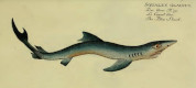


Hemipristis intermedia
Cicimurri, Ebersole, Stringer, Starnes & Phillips, 2025
Classification: Elasmobranchii Carcharhiniformes Hemigaleidae
Reference of the original description
Late Oligocene fishes (Chondrichthyes and Osteichthyes) from the Catahoula Formation in Wayne County, Mississippi, USA. European Journal of Taxonomy, 984(1), 1–131
Late Oligocene fishes (Chondrichthyes and Osteichthyes) from the Catahoula Formation in Wayne County, Mississippi, USA. European Journal of Taxonomy, 984(1), 1–131
Synonyms / new combinations and misspellings
Hemipristis cf. intermedia
Hemipristis cf. intermedia
Types
Hemipristis intermedia
Holotype: SC: 2013.28.73; Paratype: MMNS: VP-12037; MMNS: VP-12036; SC: 2013.28.80;
Hemipristis intermedia
Holotype: SC: 2013.28.73; Paratype: MMNS: VP-12037; MMNS: VP-12036; SC: 2013.28.80;
Description:
Citation: Hemipristis intermedia Cicimurri, Ebersole, Stringer, Starnes & Phillips, 2025: In: Database of fossil elasmobranch teeth www.shark-references.com, World Wide Web electronic publication, Version 07/2026
Please send your images of "Hemipristis intermedia" to info@shark-references.com
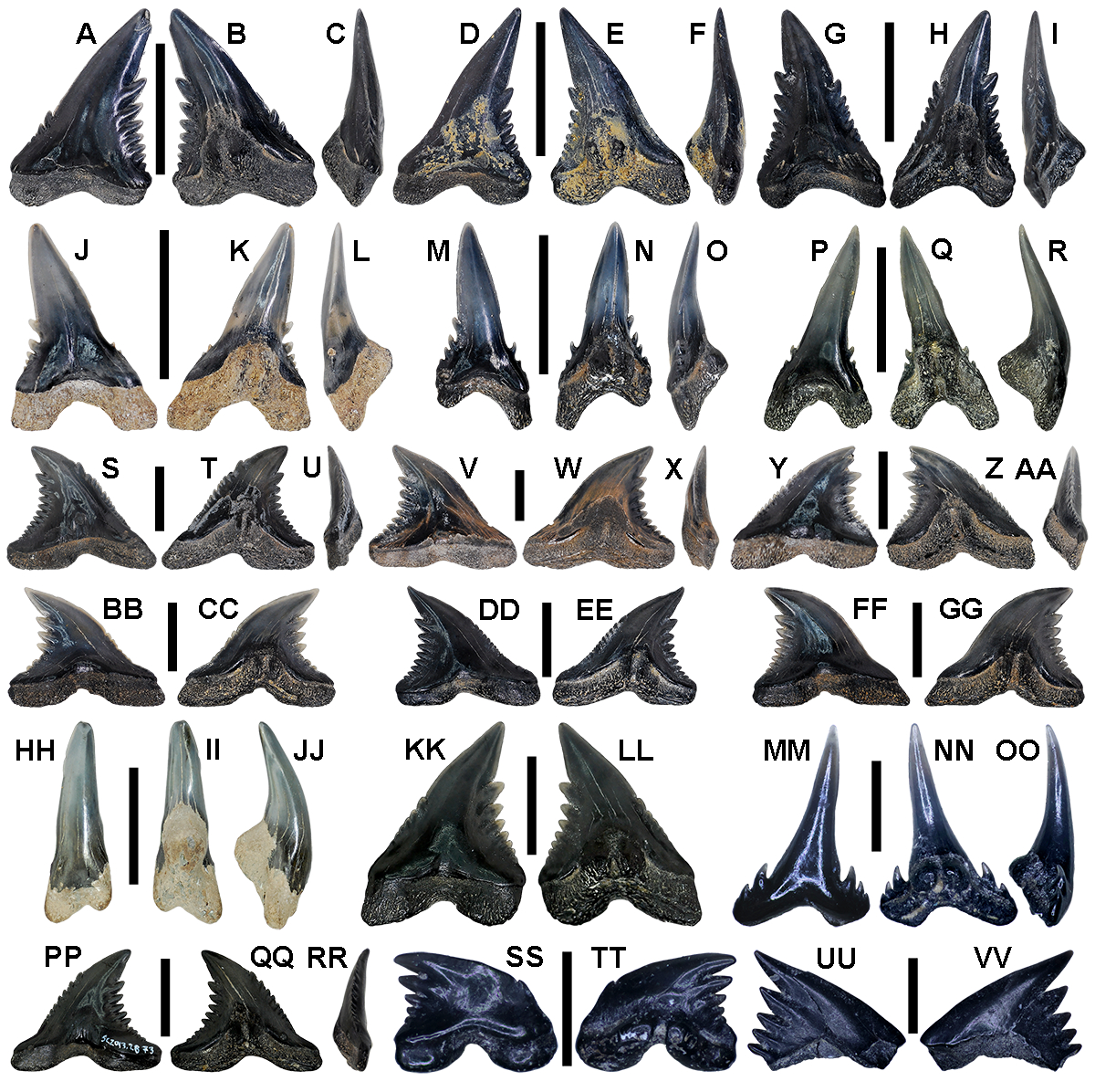
Hemipristis intermedia Cicimurri, Ebersole, Stringer, Starnes & Phillips, 2025; Fig. 6. Hemipristis intermedia sp. nov., teeth. A–C. MMNS VP-12035, lower left lateral tooth. A. Labial view. B. Lingual view. C. Mesial view. D–F. MMNS VP-12036 (paratype), lower left lateral tooth. D. Labial view. E. Lingual view. F. Mesial view. G–I. MMNS VP-12037 (paratype), upper left anterior tooth. G. Labial view. H. Lingual view. I. Mesial view. J–L. MMNS VP-12038, lower right anterolateral tooth. J. Labial view. K. Lingual view. L. Mesial view. M–O. MMNS VP-12039, lower right anterior tooth. M. Labial view. N. Lingual view. O. Mesial view. P–R. SC2013.28.80 (paratype), lower left anterior tooth. P. Labial view. Q. Lingual view. R. Mesial view. S–U. MMNS VP-12040, upper left lateral tooth. S. Labial view. T. Lingual view. U. Mesial view. V–X. MMNS VP-12042, upper left lateral tooth. V. Labial view. W. Lingual view. X. Mesial view. Y–AA. MMNS VP-12043, upper right lateral tooth. Y. Labial view. Z. Lingual view. AA. Mesial view. BB–CC. MMNS VP- 12044, upper left lateral tooth. BB. Labial view. CC. Lingual view. DD–EE. MMNS VP-12045, upper left lateral tooth. DD. Labial view. EE. Lingual view. FF–GG. MMNS VP-12046, upper left lateral tooth. FF. Labial view. GG. Lingual view. HH–JJ. MMNS VP-6625, lower left symphyseal tooth. HH. Labial view. II. Lingual view. JJ. Mesial view. KK–LL. SC2013.28.90, upper right lateral tooth. KK. Labial view. LL. Lingual view. MM–OO. MMNS VP-7604, juvenile lower left anterior tooth. MM. Labial view. NN. Lingual view. OO. Mesial view. PP–RR. SC2013.28.73 (holotype), upper right lateral tooth. PP. Labial view. QQ. Lingual view. RR. Mesial view. SS–TT. SC2013.28.103, juvenile upper left postero-lateral tooth. SS. Labial view. TT. Lingual view. UU–VV. SC2013.28.104, juvenile upper left lateral tooth. UU. Labial view. VV. Lingual view. Scale bars: A–U, HH–JJ = 1 cm; MM–OO = 2 mm; V–GG, KK–LL, PP–QQ = 5 mm; RR–UU = 1 mm. From Cicimurri et al. (2025)
Hemipristis intermedia Cicimurri, Ebersole, Stringer, Starnes & Phillips, 2025; Fig. 6. Hemipristis intermedia sp. nov., teeth. A–C. MMNS VP-12035, lower left lateral tooth. A. Labial view. B. Lingual view. C. Mesial view. D–F. MMNS VP-12036 (paratype), lower left lateral tooth. D. Labial view. E. Lingual view. F. Mesial view. G–I. MMNS VP-12037 (paratype), upper left anterior tooth. G. Labial view. H. Lingual view. I. Mesial view. J–L. MMNS VP-12038, lower right anterolateral tooth. J. Labial view. K. Lingual view. L. Mesial view. M–O. MMNS VP-12039, lower right anterior tooth. M. Labial view. N. Lingual view. O. Mesial view. P–R. SC2013.28.80 (paratype), lower left anterior tooth. P. Labial view. Q. Lingual view. R. Mesial view. S–U. MMNS VP-12040, upper left lateral tooth. S. Labial view. T. Lingual view. U. Mesial view. V–X. MMNS VP-12042, upper left lateral tooth. V. Labial view. W. Lingual view. X. Mesial view. Y–AA. MMNS VP-12043, upper right lateral tooth. Y. Labial view. Z. Lingual view. AA. Mesial view. BB–CC. MMNS VP- 12044, upper left lateral tooth. BB. Labial view. CC. Lingual view. DD–EE. MMNS VP-12045, upper left lateral tooth. DD. Labial view. EE. Lingual view. FF–GG. MMNS VP-12046, upper left lateral tooth. FF. Labial view. GG. Lingual view. HH–JJ. MMNS VP-6625, lower left symphyseal tooth. HH. Labial view. II. Lingual view. JJ. Mesial view. KK–LL. SC2013.28.90, upper right lateral tooth. KK. Labial view. LL. Lingual view. MM–OO. MMNS VP-7604, juvenile lower left anterior tooth. MM. Labial view. NN. Lingual view. OO. Mesial view. PP–RR. SC2013.28.73 (holotype), upper right lateral tooth. PP. Labial view. QQ. Lingual view. RR. Mesial view. SS–TT. SC2013.28.103, juvenile upper left postero-lateral tooth. SS. Labial view. TT. Lingual view. UU–VV. SC2013.28.104, juvenile upper left lateral tooth. UU. Labial view. VV. Lingual view. Scale bars: A–U, HH–JJ = 1 cm; MM–OO = 2 mm; V–GG, KK–LL, PP–QQ = 5 mm; RR–UU = 1 mm. From Cicimurri et al. (2025)
Description
Original diagnosis after Cicimurri, Ebersole, Stringer, Starnes & Phillips (2025) p. 18-19 [34348]: Upper lateral teeth are the most common tooth morphology represented, and because they are more diagnostic than the lower teeth, they are utilized herein to diagnose the species. Upper lateral teeth measure up to 2.5 cm in width (mesio-distal) and 2 cm in height (apico-basal). These teeth have a broad, triangular crown and distally directed main cusp. The mesial cutting edge may be smooth or bear up to ten denticles and the distal cutting edge up to 12 denticles, with denticles on both edges increasing in size towards the apex. A smooth-edged cusp constitutes the apical 30%–40% of the crown. Of the fossil Hemipristisspecies we consider valid, the upper lateral teeth of H. intermedia sp. nov. differ from those of the Eocene H. curvatus Dames, 1883 by attaining larger overall sizes (2.5 cm wide by 2.2 cm high for H. intermedia vs 1.5 cm and 1.1 cm, respectively for H. curvatus), by having more mesial and distal denticles (up to four and eight, respectively for H. curvatus, and up to 10 and 11, respectively, for H. intermedia), and by having mesial denticles that extend higher on the crown (two-thirds the crown height vs. one-half the crown height on H. curvatus). Hemipristis intermedia sp. nov. upper lateral teeth differ from those of the Miocene to Early Pleistocene H. serra Agassiz, 1835 by attaining smaller overall sizes (3.7 cm wide and 3.6 cm height for H. serra), by having fewer distal denticles (up to 20 have been observed on H. serra), and by having denticles that do not extent as close to the apex, resulting in a cusp that represents more than 20% of the crown height (as opposed to 10% in H. serra). These teeth are differentiated from those of the Rupelian H. tanakai Tomita, Yabumoto & Kuga, 2023 by having more than five mesial denticles (the maximum number reported for H. tanakai), and the apical-most mesial and distal denticles are of nearly equal height on the crown (whereas the distal denticle is generally higher in H. tanakai). Finally, the upper lateral teeth of Hemipristis intermedia sp. nov. differ from those of the extant H. elongata (Klunzinger, 1871) by being mesio-distally wider, by having more conspicuous denticles, and by having a more convex upper one-half of the mesial crown edge. The number of tooth denticles of Hemipristis intermedia is greater than the maximum occurring on H. curvatus teeth but less than the maximum number known for the Miocene to Early Pleistocene H. serra Agassiz, 1835. The proportion of cusp to total crown height is less than in H. curvatus but greater than in H. serra. The recently named H. tanakai (Tomita et al. 2023) is considered herein as a nomen dubium (see below), but the tooth size of that taxon overlaps with those of H. serra and H. intermedia. Additionally, only five mesial denticles occur in H. tanakai and the mesial denticles extend closer to the crown apex compared to the distal edge.
Original diagnosis after Cicimurri, Ebersole, Stringer, Starnes & Phillips (2025) p. 18-19 [34348]: Upper lateral teeth are the most common tooth morphology represented, and because they are more diagnostic than the lower teeth, they are utilized herein to diagnose the species. Upper lateral teeth measure up to 2.5 cm in width (mesio-distal) and 2 cm in height (apico-basal). These teeth have a broad, triangular crown and distally directed main cusp. The mesial cutting edge may be smooth or bear up to ten denticles and the distal cutting edge up to 12 denticles, with denticles on both edges increasing in size towards the apex. A smooth-edged cusp constitutes the apical 30%–40% of the crown. Of the fossil Hemipristisspecies we consider valid, the upper lateral teeth of H. intermedia sp. nov. differ from those of the Eocene H. curvatus Dames, 1883 by attaining larger overall sizes (2.5 cm wide by 2.2 cm high for H. intermedia vs 1.5 cm and 1.1 cm, respectively for H. curvatus), by having more mesial and distal denticles (up to four and eight, respectively for H. curvatus, and up to 10 and 11, respectively, for H. intermedia), and by having mesial denticles that extend higher on the crown (two-thirds the crown height vs. one-half the crown height on H. curvatus). Hemipristis intermedia sp. nov. upper lateral teeth differ from those of the Miocene to Early Pleistocene H. serra Agassiz, 1835 by attaining smaller overall sizes (3.7 cm wide and 3.6 cm height for H. serra), by having fewer distal denticles (up to 20 have been observed on H. serra), and by having denticles that do not extent as close to the apex, resulting in a cusp that represents more than 20% of the crown height (as opposed to 10% in H. serra). These teeth are differentiated from those of the Rupelian H. tanakai Tomita, Yabumoto & Kuga, 2023 by having more than five mesial denticles (the maximum number reported for H. tanakai), and the apical-most mesial and distal denticles are of nearly equal height on the crown (whereas the distal denticle is generally higher in H. tanakai). Finally, the upper lateral teeth of Hemipristis intermedia sp. nov. differ from those of the extant H. elongata (Klunzinger, 1871) by being mesio-distally wider, by having more conspicuous denticles, and by having a more convex upper one-half of the mesial crown edge. The number of tooth denticles of Hemipristis intermedia is greater than the maximum occurring on H. curvatus teeth but less than the maximum number known for the Miocene to Early Pleistocene H. serra Agassiz, 1835. The proportion of cusp to total crown height is less than in H. curvatus but greater than in H. serra. The recently named H. tanakai (Tomita et al. 2023) is considered herein as a nomen dubium (see below), but the tooth size of that taxon overlaps with those of H. serra and H. intermedia. Additionally, only five mesial denticles occur in H. tanakai and the mesial denticles extend closer to the crown apex compared to the distal edge.
Remarks
shark-references Species-ID=17561
shark-references Species-ID=17561
References


Additional records of Paleogene fishes (Chondrichthyes and Osteichthyes) from Alabama, USA. Acta Geologica Polonica, 75(4), Article e60
DOI: 10.24425/agp.2025.155954
Late Oligocene fishes (Chondrichthyes and Osteichthyes) from the Catahoula Formation in Wayne County, Mississippi, USA. European Journal of Taxonomy, 984(1), 1–131
DOI: 10.5852/ejt.2025.984.2851