


Oligoraja pristina
Reinecke, 2015
Classification: Elasmobranchii Rajiformes Rajidae
Reference of the original description
Batoids (Rajiformes, Torpediniformes, Myliobatiformes) from the Sülstorf Beds (Chattian, Late Oligocene) of Mecklenburg, northeastern Germany: a revision and description of three new species. Palaeovertebrata, 39, Article e2
Batoids (Rajiformes, Torpediniformes, Myliobatiformes) from the Sülstorf Beds (Chattian, Late Oligocene) of Mecklenburg, northeastern Germany: a revision and description of three new species. Palaeovertebrata, 39, Article e2
Types
Oligoraja pristina
Holotype: SMF(fossil): P 9849; Paratype: SMF(fossil): P 9850; SMF(fossil): P 9851; SMF(fossil): P 9852; SMF(fossil): P 9853; SMF(fossil): P 9854; SMF(fossil): P 9855; SMF(fossil): P 9856; SMF(fossil): P 9857; SMF(fossil): P 9858; SMF(fossil): P 9859;
Oligoraja pristina
Holotype: SMF(fossil): P 9849; Paratype: SMF(fossil): P 9850; SMF(fossil): P 9851; SMF(fossil): P 9852; SMF(fossil): P 9853; SMF(fossil): P 9854; SMF(fossil): P 9855; SMF(fossil): P 9856; SMF(fossil): P 9857; SMF(fossil): P 9858; SMF(fossil): P 9859;
Description:
Citation: Oligoraja pristina Reinecke, 2015: In: Database of fossil elasmobranch teeth www.shark-references.com, World Wide Web electronic publication, Version 07/2026
Please send your images of "Oligoraja pristina" to info@shark-references.com
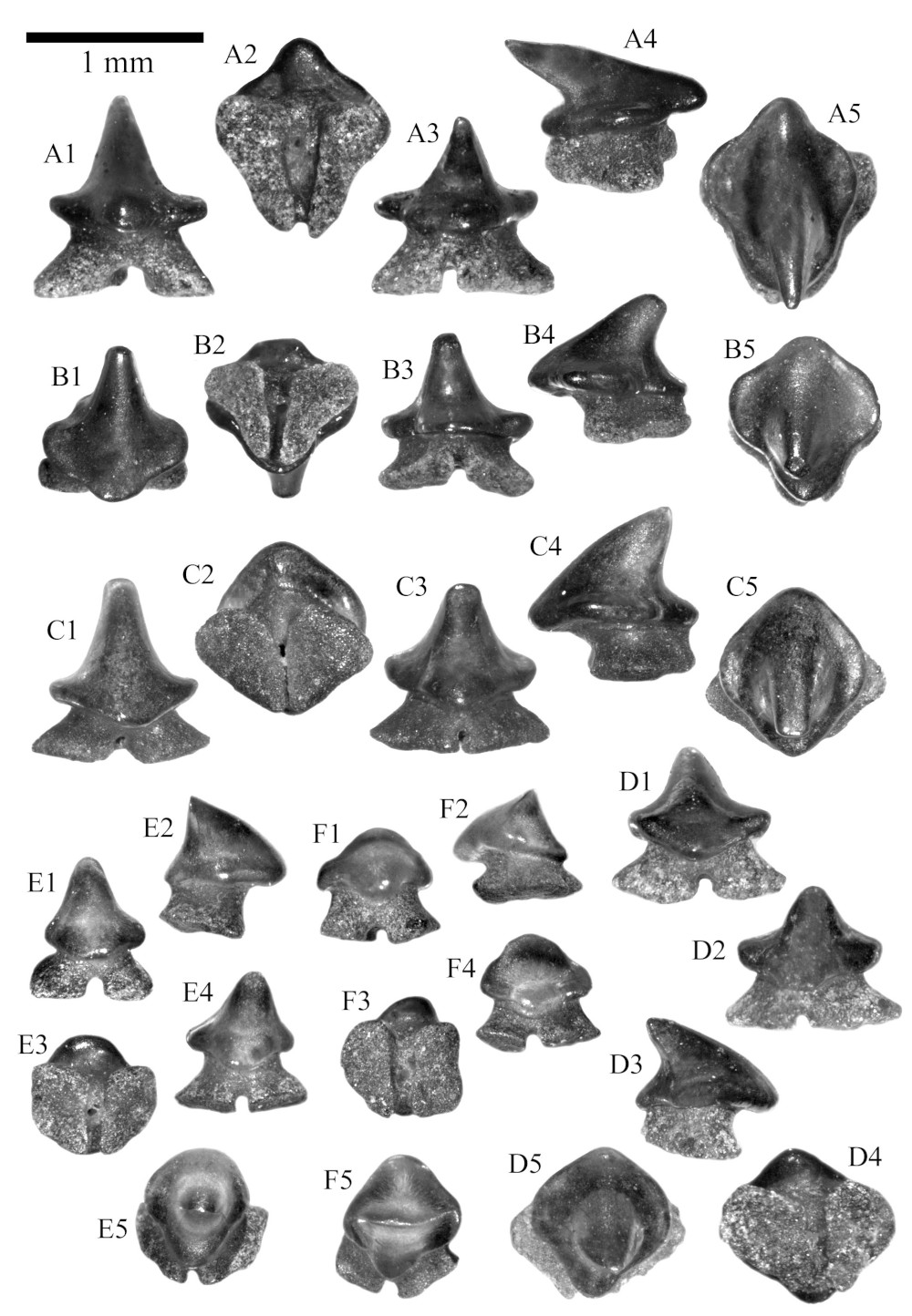
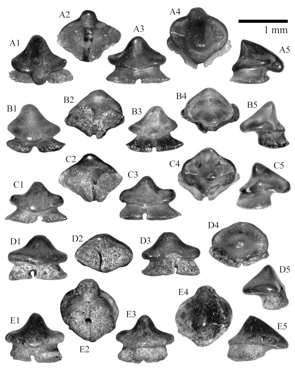
Oligoraja pristina sp. nov., Sülstorf Beds, Chattian, Kobrow, Mecklenburg-Vorpommern, Germany. A, anterior tooth of a male, H = 0.9 mm, W = 1.0 mm, L = 1.1 mm, A1 labial view, A2 basal view, A3 lingual view, A4 profile view, A5 occlusal view, SMF P 9849, holotype. B, anterior tooth of a male, H = 0.98 mm, W = 0.96 mm, L = 1.04 mm, B1 labial view, B2 basal view, B3 lingual view, B4 profile view, B5 occlusal view, SMF P 9850, paratype 1. C, anterior tooth of a male, H = 1.11 mm, W = 1.16 mm, L = 1.12 mm, C1 labial view, C2 basal view, C3 lingual view, C4 profile view, C5 occlusal view, SMF P 9851, paratype 2. D, anterior tooth of a male, H = 0.8 mm, W = 1.0 mm, L = 0.8 mm, D1 labial view, D2 lingual view, D3 profile view, D4 basal view, D5 occlusal view, SMF P 9852, paratype 3. E, anterior tooth of ? juvenile male, H = 0.8 mm, W = 0.8 mm, L = 0.7 mm, E1 labial view, E2 profile view, E3 basal view, E4 lingual view, E5 occlusal view, SMF P 9853, paratype 4. F, anterior tooth of ? juvenile male, H = 0.7 mm, W = 0.7 mm, L = 0.8 mm, F1 labial view, F2 profile view, F3 basal view, F4 lingual view, F5 occlusal view, SMF P 9854, paratype 5. © Reinecke (2015)
Oligoraja pristina sp. nov., Sülstorf Beds, Chattian, Kobrow, Mecklenburg-Vorpommern, Germany. A, anterior tooth of a male, H = 0.9 mm, W = 1.0 mm, L = 1.1 mm, A1 labial view, A2 basal view, A3 lingual view, A4 profile view, A5 occlusal view, SMF P 9849, holotype. B, anterior tooth of a male, H = 0.98 mm, W = 0.96 mm, L = 1.04 mm, B1 labial view, B2 basal view, B3 lingual view, B4 profile view, B5 occlusal view, SMF P 9850, paratype 1. C, anterior tooth of a male, H = 1.11 mm, W = 1.16 mm, L = 1.12 mm, C1 labial view, C2 basal view, C3 lingual view, C4 profile view, C5 occlusal view, SMF P 9851, paratype 2. D, anterior tooth of a male, H = 0.8 mm, W = 1.0 mm, L = 0.8 mm, D1 labial view, D2 lingual view, D3 profile view, D4 basal view, D5 occlusal view, SMF P 9852, paratype 3. E, anterior tooth of ? juvenile male, H = 0.8 mm, W = 0.8 mm, L = 0.7 mm, E1 labial view, E2 profile view, E3 basal view, E4 lingual view, E5 occlusal view, SMF P 9853, paratype 4. F, anterior tooth of ? juvenile male, H = 0.7 mm, W = 0.7 mm, L = 0.8 mm, F1 labial view, F2 profile view, F3 basal view, F4 lingual view, F5 occlusal view, SMF P 9854, paratype 5. © Reinecke (2015)
Description
Original description after Reinecke (2015) p. 12-17 [22769]: This batoid species has the smallest teeth extracted from the Sülstorf Beds, as their maximum diameter is always less than 1.4 mm. Our material can be divided into two different morphological groups. About two dozen specimens, displaying minor dimensional variation, show a cuspidate crown, set on a stocky, low root (Figs. 9A-D). The bulk of the material has lower crowns that are almost equidimensional (Fig. 10) and shows a continuous, though minor variation of height-to-width and width-to-length ratios which may be indicative of gradient heterodonty. A few smaller specimens with apically rounded, rather low cusps are intermediate between the two morphs (Figs. 9E-F). While the cuspidate, higher-crowned teeth likely represent anterior jaw positions of male individuals, the lower-crowned teeth are possibly from lateral files of male dentitions and from female dentitions. The smallest, cuspidate specimens, measuring less than 1 mm in maximum diameter (Figs. 9E-F), may be considered as anterior teeth of juvenile/ subadult males. The cuspidate teeth have a symmetrical crown with a strong, lingually inclined cusp, which merges into a laterally expanded, elliptical crown basis with lobate outline in occlusal view (see Figs. 9A5, 9B5, 9C5, 9D5). Weak cutting edges, displaced to the lingual part of the cusp, extend from the apex to half of the cusp length. A labial cutting edge (labial keel) is not present. In profile view, the labial outline of the cusp is straight (Fig. 9B4), or smoothly convex (Figs. 9C4, 9D3) or slightly kinked (Fig. 9A4). The crown tip is sometimes shortened by wear (Figs. 9B4, 5). The lingual visor has a smoothly convex outline. In the holotype, the labial visor forms a prominent bulge which markedly overhangs the root (Figs. 9A2, 4, 5). The labial bulge is less prominent in other, probably more laterally placed teeth (Figs. 9B5, 9C5, 9D5). The holaulacorhize root is always lower than the crown and branches into wide root lobes which in basal direction diverge more laterally than lingually (e.g. Figs. 9A3, 4). One or two small foramina open into the wedge-shaped nutrient groove. Marginolingual foramina are not present. The submillimetric teeth with cuspidate to globular crown morphology (Figs. 9E, F) have a variably strong labial bulge, a rounded crown apex in labial/lingual view, and a transverse crest with short mesial and distal cutting edges. Lower-crowned teeth (Figs. 10A-E) have a lateral-symmetrical crown with a transverse crest that is slightly shifted in lingual direction. In labial/lingual view the transverse crest forms a well rounded cusp which merges into oblique to subhorizontal heels. In occlusal view, the crown basis shows a lobate outline which is similar to that of cuspidate teeth (see above). The lobate shape is enhanced in teeth with the labial visor forming a distinct bulge (Figs. 10A4, 10C4, 10E4) and the lingual visor a moderate uvula (Figs. 10B4, 10C4). The crown faces are always smooth. The axial regions of the labial and lingual crown face are convex, whereas the lateral regions are flat to slightly concave. Cutting edges are missing in several teeth (Figs. 10D, 10E). If short, faint cutting edges are present in the apical region, they do not extend to the lateral heels. In profile view, the outline of the labial crown face is straight to convex, and the outline of the lingual face is variably concave (Figs. 10A5, 10C5, 10D5).
Original description after Reinecke (2015) p. 12-17 [22769]: This batoid species has the smallest teeth extracted from the Sülstorf Beds, as their maximum diameter is always less than 1.4 mm. Our material can be divided into two different morphological groups. About two dozen specimens, displaying minor dimensional variation, show a cuspidate crown, set on a stocky, low root (Figs. 9A-D). The bulk of the material has lower crowns that are almost equidimensional (Fig. 10) and shows a continuous, though minor variation of height-to-width and width-to-length ratios which may be indicative of gradient heterodonty. A few smaller specimens with apically rounded, rather low cusps are intermediate between the two morphs (Figs. 9E-F). While the cuspidate, higher-crowned teeth likely represent anterior jaw positions of male individuals, the lower-crowned teeth are possibly from lateral files of male dentitions and from female dentitions. The smallest, cuspidate specimens, measuring less than 1 mm in maximum diameter (Figs. 9E-F), may be considered as anterior teeth of juvenile/ subadult males. The cuspidate teeth have a symmetrical crown with a strong, lingually inclined cusp, which merges into a laterally expanded, elliptical crown basis with lobate outline in occlusal view (see Figs. 9A5, 9B5, 9C5, 9D5). Weak cutting edges, displaced to the lingual part of the cusp, extend from the apex to half of the cusp length. A labial cutting edge (labial keel) is not present. In profile view, the labial outline of the cusp is straight (Fig. 9B4), or smoothly convex (Figs. 9C4, 9D3) or slightly kinked (Fig. 9A4). The crown tip is sometimes shortened by wear (Figs. 9B4, 5). The lingual visor has a smoothly convex outline. In the holotype, the labial visor forms a prominent bulge which markedly overhangs the root (Figs. 9A2, 4, 5). The labial bulge is less prominent in other, probably more laterally placed teeth (Figs. 9B5, 9C5, 9D5). The holaulacorhize root is always lower than the crown and branches into wide root lobes which in basal direction diverge more laterally than lingually (e.g. Figs. 9A3, 4). One or two small foramina open into the wedge-shaped nutrient groove. Marginolingual foramina are not present. The submillimetric teeth with cuspidate to globular crown morphology (Figs. 9E, F) have a variably strong labial bulge, a rounded crown apex in labial/lingual view, and a transverse crest with short mesial and distal cutting edges. Lower-crowned teeth (Figs. 10A-E) have a lateral-symmetrical crown with a transverse crest that is slightly shifted in lingual direction. In labial/lingual view the transverse crest forms a well rounded cusp which merges into oblique to subhorizontal heels. In occlusal view, the crown basis shows a lobate outline which is similar to that of cuspidate teeth (see above). The lobate shape is enhanced in teeth with the labial visor forming a distinct bulge (Figs. 10A4, 10C4, 10E4) and the lingual visor a moderate uvula (Figs. 10B4, 10C4). The crown faces are always smooth. The axial regions of the labial and lingual crown face are convex, whereas the lateral regions are flat to slightly concave. Cutting edges are missing in several teeth (Figs. 10D, 10E). If short, faint cutting edges are present in the apical region, they do not extend to the lateral heels. In profile view, the outline of the labial crown face is straight to convex, and the outline of the lingual face is variably concave (Figs. 10A5, 10C5, 10D5).
Remarks
shark-references Species-ID=14394;
type species of Oligoraja Reinecke, 2015 p. 14 [22769] by original designation (Art. 68.2 ICZN);
valid after Reinecke (2015) p. 14 [22769];
shark-references Species-ID=14394;
type species of Oligoraja Reinecke, 2015 p. 14 [22769] by original designation (Art. 68.2 ICZN);
valid after Reinecke (2015) p. 14 [22769];
Image gallery

References


Batoids (Rajiformes, Torpediniformes, Myliobatiformes) from the Sülstorf Beds (Chattian, Late Oligocene) of Mecklenburg, northeastern Germany: a revision and description of three new species. Palaeovertebrata, 39, Article e2
Batoids (Rajiformes, Torpediniformes, Myliobatiformes) from the Sülstorf Beds (Chattian, Late Oligocene) of Mecklenburg, northeastern Germany: a revision and description of three new species. Palaeovertebrata, 39, Article e2