


Torpedo chattica
Reinecke, 2015
Classification: Elasmobranchii Torpediniformes Torpedinidae
Reference of the original description
Batoids (Rajiformes, Torpediniformes, Myliobatiformes) from the Sülstorf Beds (Chattian, Late Oligocene) of Mecklenburg, northeastern Germany: a revision and description of three new species. Palaeovertebrata, 39, Article e2
Batoids (Rajiformes, Torpediniformes, Myliobatiformes) from the Sülstorf Beds (Chattian, Late Oligocene) of Mecklenburg, northeastern Germany: a revision and description of three new species. Palaeovertebrata, 39, Article e2
Types
Torpedo chattica
Holotype: SMF(fossil): P 9866; Paratype: SMF(fossil): P 9860; SMF(fossil): P 9861; SMF(fossil): P 9862; SMF(fossil): P 9863; SMF(fossil): P 9864;
Torpedo chattica
Holotype: SMF(fossil): P 9866; Paratype: SMF(fossil): P 9860; SMF(fossil): P 9861; SMF(fossil): P 9862; SMF(fossil): P 9863; SMF(fossil): P 9864;
Description:
Citation: Torpedo chattica Reinecke, 2015: In: Database of fossil elasmobranch teeth www.shark-references.com, World Wide Web electronic publication, Version 07/2026
Please send your images of "Torpedo chattica" to info@shark-references.com
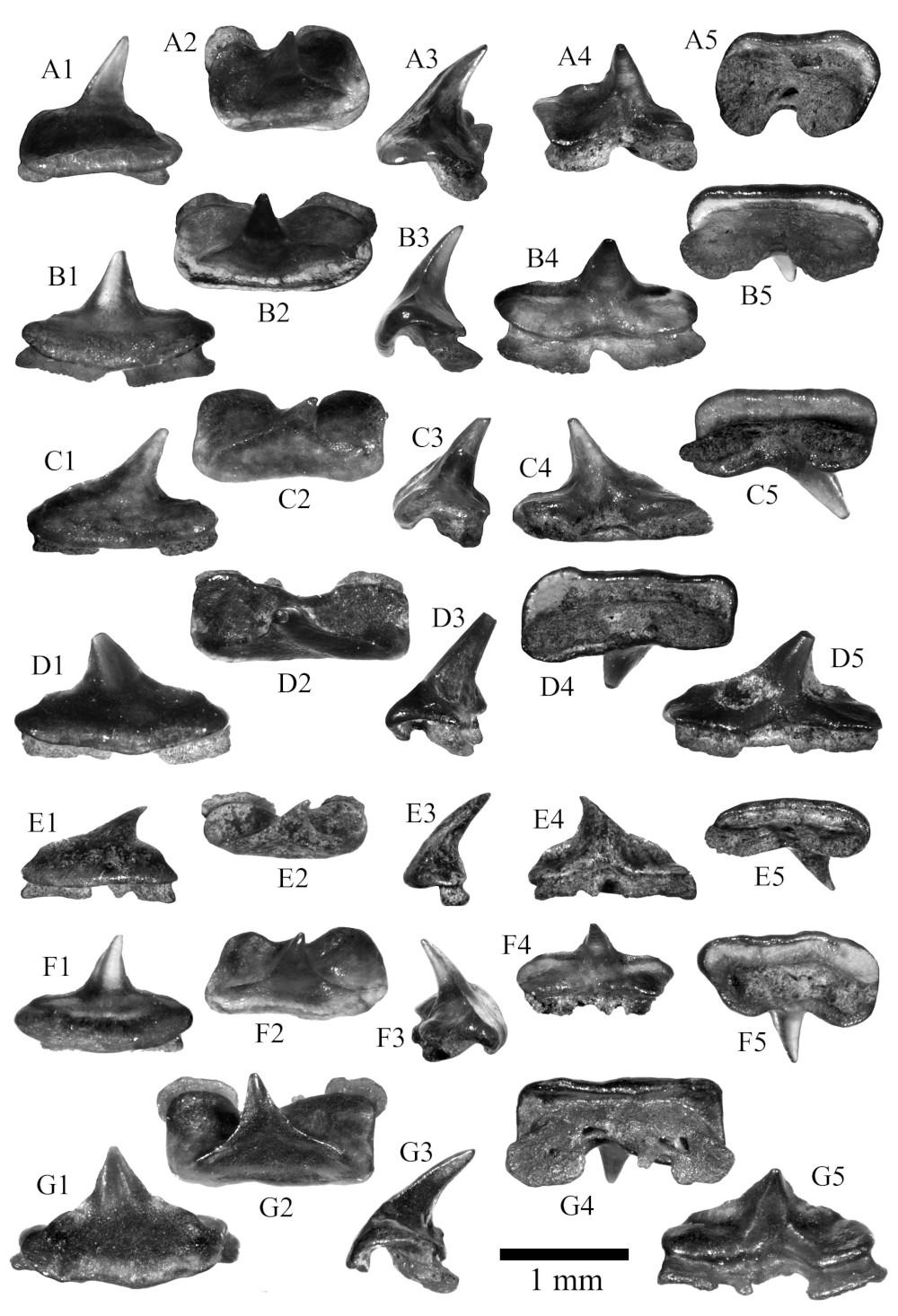
Torpedo chattica sp. nov., Sülstorf Beds, Chattian, Kobrow, Mecklenburg-Vorpommern, Germany. A, anterior tooth, H = 1.1 mm, W = 1.3 mm, L = 0.9 mm, A1 labial view, A2 occlusal view, A3 profile view, A4 lingual view, A5 basal view, SMF P 9860, paratype 1. B, anterolateral tooth, H = 1.0 mm, W = 1.6 mm, L = 0.8 mm, B1 labial view, B2 occlusal view, B3 profile view, B4 lingual view, B5 basal view, SMF P 9861, paratype 2. C, anterolateral tooth, H = 1.0 mm, W = 1.5 mm, L = 0.8 mm, C1 labial view, C2 occlusal view, C3 profile view, C4 lingual view, C5 basal view, SMF P 9862, paratype 3. D, anterolateral tooth, H = 1.0 mm, W = 1.7 mm, L = 0.8 mm, D1 labial view, D2 occlusal view, D3 profile view, D4 basal view, D5 lingual view, SMF P 9863. E, lateroposterior tooth, H = 0.7 mm, W = 1.2 mm, L = 0.5 mm, E1 labial view, E2 occlusal view, E3 profile view, E4 lingual view, E5 basal view, SMF P 9864, paratype 4. F, anterolateral tooth, H = 0.9 mm, W = 1.4 mm, L = 0.8 mm, F1 labial view, F2 occlusal view, F3 profile view, F4 lingual view, F5 basal view, SMF P 9865. G, anterolateral tooth, H = 1.3 mm, W = 1.8 mm, L = 0.9 mm, G1 labial view, G2 occlusal view, G3 profile view, G4 basal view, G5 lingual view, SMF P 9866, holotype. © Reinecke (2015)
Torpedo chattica sp. nov., Sülstorf Beds, Chattian, Kobrow, Mecklenburg-Vorpommern, Germany. A, anterior tooth, H = 1.1 mm, W = 1.3 mm, L = 0.9 mm, A1 labial view, A2 occlusal view, A3 profile view, A4 lingual view, A5 basal view, SMF P 9860, paratype 1. B, anterolateral tooth, H = 1.0 mm, W = 1.6 mm, L = 0.8 mm, B1 labial view, B2 occlusal view, B3 profile view, B4 lingual view, B5 basal view, SMF P 9861, paratype 2. C, anterolateral tooth, H = 1.0 mm, W = 1.5 mm, L = 0.8 mm, C1 labial view, C2 occlusal view, C3 profile view, C4 lingual view, C5 basal view, SMF P 9862, paratype 3. D, anterolateral tooth, H = 1.0 mm, W = 1.7 mm, L = 0.8 mm, D1 labial view, D2 occlusal view, D3 profile view, D4 basal view, D5 lingual view, SMF P 9863. E, lateroposterior tooth, H = 0.7 mm, W = 1.2 mm, L = 0.5 mm, E1 labial view, E2 occlusal view, E3 profile view, E4 lingual view, E5 basal view, SMF P 9864, paratype 4. F, anterolateral tooth, H = 0.9 mm, W = 1.4 mm, L = 0.8 mm, F1 labial view, F2 occlusal view, F3 profile view, F4 lingual view, F5 basal view, SMF P 9865. G, anterolateral tooth, H = 1.3 mm, W = 1.8 mm, L = 0.9 mm, G1 labial view, G2 occlusal view, G3 profile view, G4 basal view, G5 lingual view, SMF P 9866, holotype. © Reinecke (2015)
Description
Original description after Reinecke (2015) p. 18 [22769]: Nine torpedinid teeth, including the two specimens previously illustrated (Reinecke et al., 2005), were recovered from the Sülstorf Beds. These form a morphologically homogeneous sample which can be distinguished from all fossil and extant species for which dental characters have been illustrated. The teeth show slight differences in the width-to-length ratio of the crown basis, the cusp height and inclination of the cusp. These can be interpreted as due to a slight gradient monognathic heterodonty. One specimen (Fig. 11A) displays a noticeable crown asymmetry and rather high height-to-width ratio (1.1/1.3). It is tentatively referred to an anterior jaw position. Other specimens have a mesio-distally more elongate crown basis and are likely derived from anterolateral files (Figs. 11B-D, 11F-G), while the smallest and labio-lingually thinnest tooth, equipped with a low, hooked cusp (Fig. 11E), may represent a lateroposterior tooth. In occlusal or basal view, the crown basis of most teeth is nearly twice as wide (mesio-distally) as long (labio-lingually), and has a broadly rectangular outline with well rounded lateroanterior and lateroposterior edges. The labial visor is straight or slightly flexuous in basal view (e.g. Figs. 11B5, 11C5, 11D4). In labial view, it shows a basally convex outline (Figs. 11B1, 11C1, 11D1, 11F1) or - more rarely - an almost straight outline (Figs. 11A1, 11E1). In profile view, the labial visor forms a narrow, convex bulge (apron) which markedly overhangs the root-crown junction. The axial region of the lingual crown basis shows a pronounced depression (in occlusal view), which divides the lingual crown basis into wide mesial and distal lobes and is placed just above the nutrient groove of the root. In two teeth the axial depression is more irregular in outline, resulting in differently large mesial and distal lobes of the lingual crown basis (Figs. 11D2, D4 and 11F2, F5). The crown basis bears a moderately high, narrow cusp which is erect or distally inclined in labial view. Mesial and distal cutting edges extend from the apex (or the upper thirds of the cusp) onto the anterior region of the crown basis, but disappear before reaching the lateral crown edges. In labial view, the lateral cutting edges appear as a short, subhorizontal ridges which on one or either sides terminate in a low, broad cusplet or hump. The labial crown face is rather flat (in profile view), whereas the lingual crown face is convex at the cusp and flat to slightly concave at the basal crown lobes. The holaulacorhize root is much lower than the crown and markedly displaced in lingual direction with respect to the vertical crown axis (e.g. Figs. 11A3, 11B3). The thin root lobes are curved in lingual direction and separated by a wide nutrient groove. One large foramen occurs in the nutrient groove. The holotype (Fig. 11G) combines the typical dental characters described above, whereas the paratypes (Figs. 11A-C, E) are chosen to represent the range of morphological variation.
Original description after Reinecke (2015) p. 18 [22769]: Nine torpedinid teeth, including the two specimens previously illustrated (Reinecke et al., 2005), were recovered from the Sülstorf Beds. These form a morphologically homogeneous sample which can be distinguished from all fossil and extant species for which dental characters have been illustrated. The teeth show slight differences in the width-to-length ratio of the crown basis, the cusp height and inclination of the cusp. These can be interpreted as due to a slight gradient monognathic heterodonty. One specimen (Fig. 11A) displays a noticeable crown asymmetry and rather high height-to-width ratio (1.1/1.3). It is tentatively referred to an anterior jaw position. Other specimens have a mesio-distally more elongate crown basis and are likely derived from anterolateral files (Figs. 11B-D, 11F-G), while the smallest and labio-lingually thinnest tooth, equipped with a low, hooked cusp (Fig. 11E), may represent a lateroposterior tooth. In occlusal or basal view, the crown basis of most teeth is nearly twice as wide (mesio-distally) as long (labio-lingually), and has a broadly rectangular outline with well rounded lateroanterior and lateroposterior edges. The labial visor is straight or slightly flexuous in basal view (e.g. Figs. 11B5, 11C5, 11D4). In labial view, it shows a basally convex outline (Figs. 11B1, 11C1, 11D1, 11F1) or - more rarely - an almost straight outline (Figs. 11A1, 11E1). In profile view, the labial visor forms a narrow, convex bulge (apron) which markedly overhangs the root-crown junction. The axial region of the lingual crown basis shows a pronounced depression (in occlusal view), which divides the lingual crown basis into wide mesial and distal lobes and is placed just above the nutrient groove of the root. In two teeth the axial depression is more irregular in outline, resulting in differently large mesial and distal lobes of the lingual crown basis (Figs. 11D2, D4 and 11F2, F5). The crown basis bears a moderately high, narrow cusp which is erect or distally inclined in labial view. Mesial and distal cutting edges extend from the apex (or the upper thirds of the cusp) onto the anterior region of the crown basis, but disappear before reaching the lateral crown edges. In labial view, the lateral cutting edges appear as a short, subhorizontal ridges which on one or either sides terminate in a low, broad cusplet or hump. The labial crown face is rather flat (in profile view), whereas the lingual crown face is convex at the cusp and flat to slightly concave at the basal crown lobes. The holaulacorhize root is much lower than the crown and markedly displaced in lingual direction with respect to the vertical crown axis (e.g. Figs. 11A3, 11B3). The thin root lobes are curved in lingual direction and separated by a wide nutrient groove. One large foramen occurs in the nutrient groove. The holotype (Fig. 11G) combines the typical dental characters described above, whereas the paratypes (Figs. 11A-C, E) are chosen to represent the range of morphological variation.
References


Batoids (Rajiformes, Torpediniformes, Myliobatiformes) from the Sülstorf Beds (Chattian, Late Oligocene) of Mecklenburg, northeastern Germany: a revision and description of three new species. Palaeovertebrata, 39, Article e2
Batoids (Rajiformes, Torpediniformes, Myliobatiformes) from the Sülstorf Beds (Chattian, Late Oligocene) of Mecklenburg, northeastern Germany: a revision and description of three new species. Palaeovertebrata, 39, Article e2